21 Feb 2024 Newton Medal Award Meeting

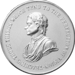
The Newton Medal is awarded every two years since 1963 by the Colour Group (Great Britain) and recognises distinguished workers in the field of colour science. We are pleased to announce that Professor John L Barbur has been selected as the recipient of the Newton Medal 2024 for his exceptional contribution to fundamental research and practical applications in colour vision, which have impacted advanced scientific understanding, public health and society.
 The Newton Medal 2024 meeting will be held on Wednesday, 21 February 2024. Professor Barbur will deliver his Newton Medal Lecture, “Variability, colour thresholds and chromatic mechanisms,” followed by the medal presentation and celebration reception.
Venue
B200, University Building at City, University of London
Programme
17:30 Welcome and introduction
17:35 Newton Lecture
18:35 Medal presentation and photos
18:40 Celebration reception with food and drinks (at Pavilion)
Professor John L Barbur: Résumé of research achievements
 John Barbur studied Physics, Optics and Visual Science at Imperial College, University of London. He has a long record of research achievements and broader impact in fundamental vision science and applied and clinical research. His early work on camouflage led to insights into the processing of luminance and colour signals with important applications in visually demanding occupations and the clinic.
John developed research instrumentation and measurement techniques to study mesopic vision, eye movements, visual search, motion and rapid flicker sensitivity, the function of the pupil response, the effects of scattered light on spatial vision, congenital and acquired colour deficiency, colour constancy and the measurement of stimulus-specific, cortical visual processing times. John’s novel tests and instrumentation led to new findings. The P_SCAN system was amongst the first developments and enabled the discovery of new components of the pupil response that require the normal processing of stimulus attributes such as colour or motion in central areas of the visual cortex. The system made possible studies of normal colour vision, congenital and acquired deficiency and ‘Blindsight’.
As a Fulbright Scholar, John worked with colleagues at the Center for Visual Science at the University of Rochester, New York. This work led to studies on mesopic optimisation of visual efficiency, medical aspects of fitness to drive, minimisation of ‘glare’ in lighting installations and mesopic optimisation of lighting for residential streets. These studies supported the development of several Advanced Vision and Optometric Tests (AVOT), initially for research and later for more precise assessment of vision and severity of vision loss in visually demanding working environments such as aviation, seafaring and rail transport. The tests include the Colour Assessment and Diagnosis (CAD) test, which is now used throughout the world to assess pilots, firefighters, seafarers, police officers and air traffic controllers. Other AVOT tests are also being used to detect changes in oculomotor responses in degenerative disorders of the brain and early-stage degenerative disorders of the retina and to evaluate the outcome of treatments in genetic clinical trials.
Current projects include the development of specific tests for non-invasive assessment of retinal function and validating a highly sensitive and specific colour vision screener with direct impact in schools, occupations and the clinic. The many research investigations which John and his colleagues carried out in the past led to the formation of the Centre for Applied Vision Research (formally known as the Applied Vision Research Centre), which is now recognised internationally as a leading centre in visual psychophysics, ocular optics, occupational vision and colour research.
Newton Medal 2024 Lecture: ‘Variability, colour thresholds and chromatic mechanisms’
Abstract
It is not unreasonable to expect some differences in human visual perception given the innumerable differences between human eyes, and the role inferential brain mechanisms play in generating a representation of what is most likely to be present in the visual scene. Many inter subject differences remain poorly understood, but the advantages of ‘seeing’ things ‘the same way’, often makes us forget about the real differences.
Fortunately, some limits are imposed on what we all see by the largely invariant properties of the five, spectrally distinct sensors in the eye. What we know for sure is that much of the spatial information carried in modulations of intensity and spectral content in retinal images is captured in four, retinal photoreceptor pigments. This information is then ‘condensed’ in the retina to provide us with efficient encoding of edges and contours defined by either luminance contrast or by spatial variations in the spectral content of the image. The latter form the basis for the red / green and yellow / blue chromatic signals. Other mechanisms which operate best at lower light levels and rely on both excitatory and inhibitory interaction of signals from rods and cones to make it all work, enable the human eye to function over an enviable range of light levels. In addition, the output of a small population of ganglion cells with large receptive fields, which receive inputs from virtually all photoceptors, is also modulated by melanopsin, a fifth photopigment located within the cell, which responds at higher light levels. These ganglion cells form a separate vision channel which may play an important role in sensing the amount of light in the visual scene and may also contribute to our ability to judge the level of ambient light. The effectiveness of these different signals generated in the retina varies greatly with changes in stimulus conditions, but all these signals can contribute directly and can affect the appearance of the objects we see.
In this lecture I shall make use mostly of colour vision and shall focus on how either changes in or the absence of one chromatic mechanism can influence the effectiveness of the remaining vision channels and how this can affect our functional vision and the ability to carry out visual tasks, including those involved in colour assessment.
In order to understand the results of colour assessment tests when plotted in colour spaces defined for subjects with normal colour vision, we developed a new model of colour discrimination to derive predictions of colour thresholds in subjects with variant cone pigments and to describe how photopic and scotopic luminance signals can affect colour assessment outcomes under conditions that are deemed to be photopically isoluminant for subjects with normal colour vision. The model also predicts accurately how the selective absorption of short wavelength light by the lens and the macular pigment in the eye can affect the orientation and size of colour threshold ellipses. Equally important, a default outcome of the model is that colour threshold contours can be plotted as circles to produce a uniform colour space. When the combined colour threshold signal strength needed to just see a colour difference between the stimulus and its adjacent background is no longer invariant with the direction of chromatic change, the results are indicative of either congenital or acquired colour deficiency.
In addition to normal colour vision and congenital colour deficiency, I shall also be presenting results from clinical studies to illustrate how functional vision can change when the binocular summation and inhibition of signals no longer functions normally or when the brightness channel no longer signals the presence of light.
